Medical depiction of metastases illustrating the spread of cancer cells throughout the body.
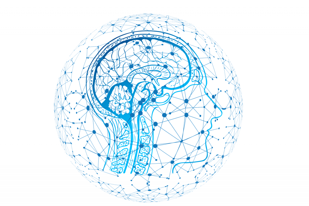
Metastases are settlements of other tumors growing in the body. They occur in the brain or spinal cord, among others.
Symptoms depend on the location of the metastasis (site of the brain) and can range from mild headaches to neurological failure or seizures. Very often, metastases in the brain or spinal cord are diagnosed only as an incidental finding during routine staging because patients do not show symptoms. Thin-slice MRI images (with a layer thickness of 1 mm or less and with contrast media, native, T2 and CISS special sequences) are then required to visualize the tumor as well as neuroanatomical structures.
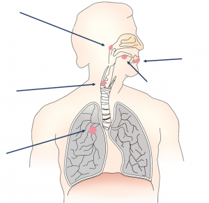
Patients with active tumors must undergo regular full-body examinations, known as staging. These examinations are used to determine how far the (malignant) tumor has spread. CT images of the thorax (= chest), abdomen and usually also the skull are taken with contrast media. If a brain or spinal metastasis is suspected, the medical imaging is complemented with an MRI (“tube”).
The treatment of metastases in the area of the brain and spinal cord is always interdisciplinary – i. e. in cooperation with other specialist clinics and in close coordination with the doctor colleagues treating the primary tumor. Depending on the treatment response of the primary tumor, a decision is then made on the therapy of the metastasis or metastases. Inside the brain or spinal cord, metastases can also be removed with minimally invasive technique. This allows rapid follow-up treatment of the primary tumor, either by chemotherapy or radiation of the resection area
(= the site where the metastasis was removed).
Metastases are settlements of other tumors growing in the body. They occur in the brain or spinal cord, among others.
Symptoms depend on the location of the metastasis (site of the brain) and can range from mild headaches to neurological failure or seizures. Very often, metastases in the brain or spinal cord are diagnosed only as an incidental finding during routine staging because patients do not show symptoms. Thin-slice MRI images (with a layer thickness of 1 mm or less
and with contrast media, native, T2 and CISS special sequences) are then required to visualize the tumor as well as neuroanatomical structures.
Patients with active tumors must undergo regular full-body examinations, known as staging. These examinations are used to determine how far the (malignant) tumor has spread. CT images of the thorax (= chest), abdomen and usually also the skull are taken with contrast media. If a brain or spinal metastasis is suspected, the medical imaging is complemented with an MRI (“tube”).
The treatment of metastases in the area of the brain and spinal cord is always interdisciplinary – i. e. in cooperation with other specialist clinics and in close coordination with the doctor colleagues treating the primary tumor. Depending on the treatment response of the primary tumor, a decision is then made on the therapy of the metastasis or metastases. Inside the brain or spinal cord, metastases can also be removed with minimally invasive technique. This allows rapid follow-up treatment of the primary tumor, either by chemotherapy or radiation of the resection area (= the site where the metastasis was removed).
Understanding Metastases
Understanding Metastases
Surgical Planning with VR and 3D Technology
Surgical Planning with VR and 3D Technology
Request for Treatment
Request for Treatment