Minimally Invasive Neurosurgery
Minimally Invasive Neurosurgery
With the term minimally invasive neurosurgery, Prof. Feigl does not simply describe the size of the surgical approaches. Instead, it refers to a surgical treatment concept in which the neurosurgical approaches (“incision”) are individually planned and adapted to the patient’s specific situation. The surgical accesses are not simply made as small as possible, but as gently as possible and therefore not larger than necessary for operational therapy. This results in minimization of the operative trauma, i. e. the planned “accompanying injury” caused by the procedure such as skin incision, openings of the skull etc.
This has the great advantage for patients that their wounds heal faster, their hospital stay is up to five days shorter, and they can return to their normal lives more quickly. Since the patients’ wounds are almost exclusively glued by Prof. Feigl, showering and hair washing is already possible the day after surgery.
Also, it saves the patient from the often very painful removal of stitches.
Minimally invasive procedures at the skull base
Minimally invasive procedures at the skull base
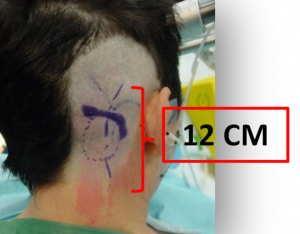
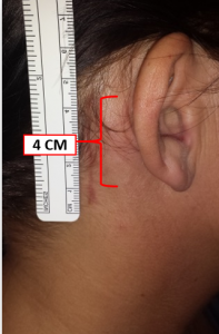
Prof. Feigl is one of only a few specialists in German-speaking countries for whom minimally invasive neurosurgery is the standard for all surgical procedures on the brain and spine. Most procedures at the base of the skull require only a minimal shave of the hair and a small craniotomy (= opening of the skull) of only about 2-3 cm, even in large tumors, due to the use of special instruments.
In addition, a 3D (three-dimensional) and VR (virtual reality) representation is used for planning of the procedures as well as for patient education. For exact positioning of the craniotomy during surgery, neuronavigation (“GPS for the brain”) based on magnetic resonance imaging and computed tomography (here: MRI or CT imaging) is always used in conjunction with the 3D and VR system.
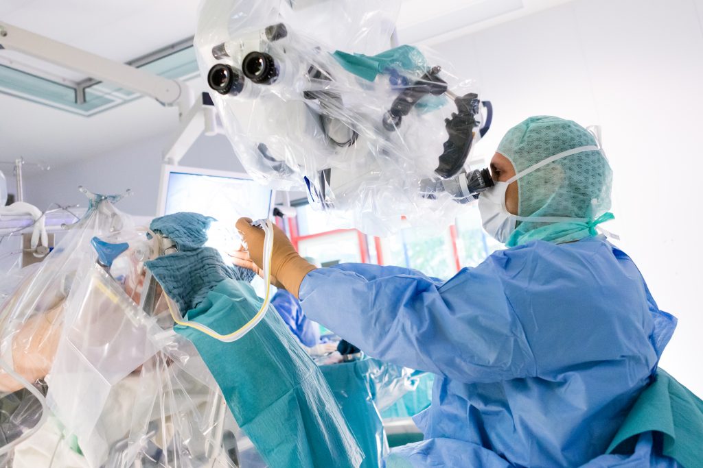
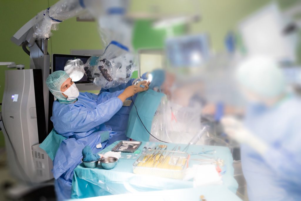
Furthermore, this treatment concept requires the use of special small microsurgical instruments, so-called “tube shaft instruments”, and a high-resolution microscope with up to 35 times magnification. In addition, fade-in of the derived nerve flows (= representation of brain nerves under the surgical microscope with fading in of the derived brain waves, which the neurosurgeons can monitor during surgery) as well as the use of a neuroendoscope with HD resolution are indispensable.
Prof. Feigl is one of only a few specialists in German-speaking countries for whom minimally invasive neurosurgery is the standard for all surgical procedures on the brain and spine. Most procedures at the base of the skull require only a minimal shave of the hair and a small craniotomy (= opening of the skull) of only about 2-3 cm, even in large tumors, due to the use of special instruments.
In addition, a 3D (three-dimensional) and VR (virtual reality) representation is used for planning of the procedures as well as for patient education. For exact positioning of the craniotomy during surgery, neuronavigation (“GPS for the brain”) based on magnetic resonance imaging and computed tomography (here: MRI or CT imaging) is always used in conjunction with the 3D and VR system.
Furthermore, this treatment concept requires the use of special small microsurgical instruments, so-called “tube shaft instruments”, and a high-resolution microscope with up to 35 times magnification.
In addition, fade-in of the derived nerve flows (= representation of brain nerves under the surgical microscope with fading in of the derived brain waves, which the neurosurgeons can monitor during surgery) as well as the use of a neuroendoscope with HD resolution are indispensable.