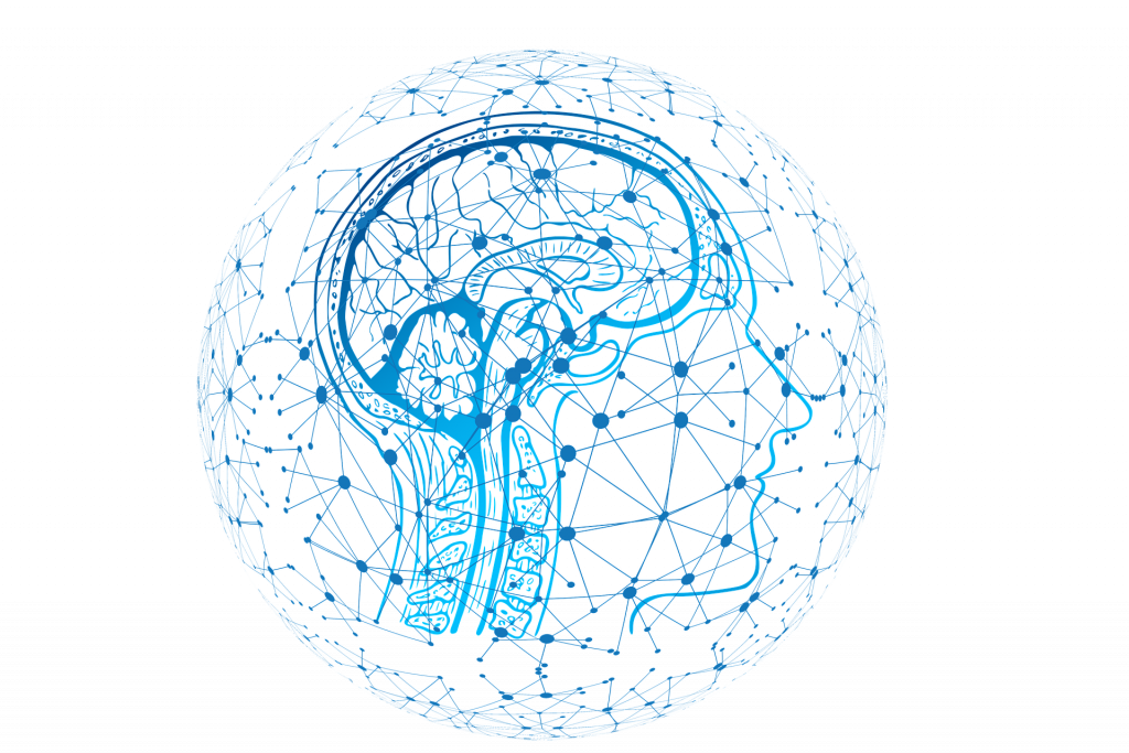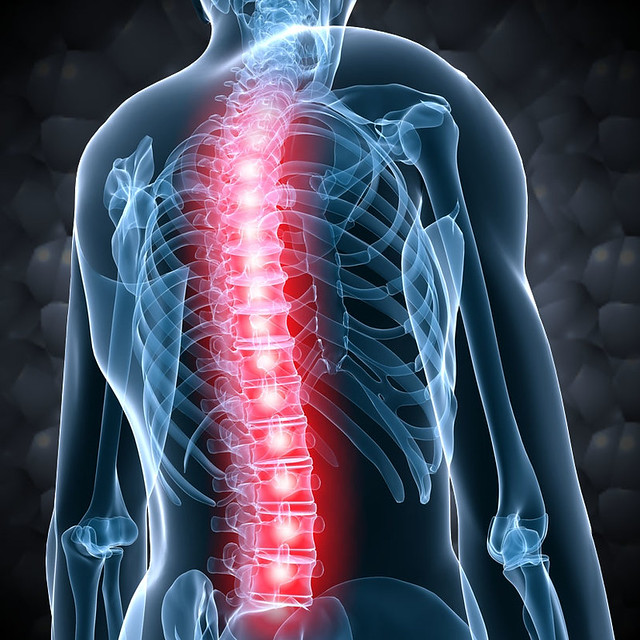
Many tumors that occur in the brain may also appear in the spinal cord, which is part of the central nervous system. These range from (malignant) gliomas to (mostly benign) meningiomas and vascular malformations (arteriovenous (AV) fistula = short circuit between artery and vein; malformations of blood or lymph vessels).
The symptoms vary depending on the exact location in the area of the spine, i. e. whether a lesion (= increase in volume inside the body) occurs in the area of the cervical, thoracic or lumbar spine. However, tumors of the spinal cord are usually noticed due to neurological deficits such as sensation or gait disorders and paralysis.
In the case of the very rare AV fistula, clinical symptoms such as sense disturbances and paralysis are only slowly developing due to the swelling of the spinal cord. The AV fistula is often detected very late as the symptoms are initially attributed to degenerative diseases of the spine and nervous system.
In case of tumors of the spine and spinal cord, neuroradiological imaging
in the MRI (“tube”) must be carried out in addition to nerve current measurements (measurement of the nerve conduction velocity;
indicates the presence of nerve damage) and a detailed clinical-neurological examination. Thin-slice MRI images (with a layer thickness of 1 mm or less with contrast media, native, T2 and CISS special sequences) are required to represent the nerves.
Arteriovenous fistulas are visualized angiographically (= in terms of vascular representation) in the MRI with a DSA (Digital Subtraction Angiography) after diagnosis. The diagnostic procedure of DSA allows the subtraction (“making invisible”) of disturbing aspects in the image.
Tumors in the area of the spinal cord can initially be treated by a microsurgical procedure. In such an operation, Prof. Feigl uses minimally invasive endoscopy-supported surgical techniques. This ensures that the surgical approach is not larger than absolutely necessary, thus allowing, among other things, a faster recovery of the patient. In addition, it is possible to combine the procedure with subsequent radiation therapy or even the exclusive treatment with radiotherapy.
As with all other tumors, the treatment strategy depends primarily on the type and spread, but also on histology and tissue structure. Histology can only be obtained as part of a surgical procedure.
Treatment of spinal AV fistulas (= belonging to the spine, to the spinal cord) is either interventional – (with a catheter inserted through the groin under X-ray illumination) by bonding the short circuit between artery and vein – or surgical, if bonding is not possible. Interventional diagnostic or therapeutic procedures are usually targeted interventions on the infected tissue.
Many tumors that occur in the brain may also appear in the spinal cord, which is part of the central nervous system. These range from (malignant) gliomas to (mostly benign) meningiomas and vascular malformations (arteriovenous (AV) fistula = short circuit between artery and vein; malformations of blood or lymph vessels).
The symptoms vary depending on the exact location in the area of the spine, i. e. whether a lesion (= increase in volume inside the body) occurs in the area of the cervical, thoracic or lumbar spine. However, tumors of the spinal cord are usually noticed due to neurological deficits such as sensation or gait disorders and paralysis.
In the case of the very rare AV fistula, clinical symptoms such as sense disturbances and paralysis are only slowly developing due to the swelling of the spinal cord. The AV fistula is often detected very late as the symptoms are initially attributed to degenerative diseases of the spine and nervous system.
In case of tumors of the spine and spinal cord, neuroradiological imaging in the MRI (“tube”) must be carried out in addition to nerve current measurements (measurement of the nerve conduction velocity; indicates the presence
of nerve damage) and a detailed clinical-neurological examination. Thin-slice MRI
images (with a layer thickness of 1 mm or less with contrast media, native, T2 and CISS special sequences) are required to represent the nerves.
Arteriovenous fistulas are visualized angiographically (= in terms of vascular representation) in the MRI with a DSA (Digital Subtraction Angiography) after diagnosis.
The diagnostic procedure of DSA allows the subtraction (“making invisible”) of disturbing aspects in the image.
Tumors in the area of the spinal cord can initially be treated by a microsurgical procedure. In such an operation, Prof. Feigl uses minimally invasive endoscopy-supported surgical techniques. This ensures that the surgical approach is not larger than absolutely necessary, thus allowing, among other things, a faster recovery of the patient. In addition, it is possible to combine the procedure with subsequent radiation therapy or even the exclusive treatment with radiotherapy.
As with all other tumors, the treatment strategy depends primarily on the type and spread, but also on histology and tissue structure.
Histology can only be obtained as part of a surgical procedure.
Treatment of spinal AV fistulas (= belonging to the spine, to the spinal cord) is either interventional – (with a catheter inserted through the groin under X-ray illumination) by bonding the short circuit between artery and vein – or surgical, if bonding is not possible. Interventional diagnostic or therapeutic procedures are usually targeted interventions on the infected tissue.
Request for Treatment
Request for Treatment