Navigation and Imaging with VR and 3D Technology
Region of the Pineal Tumor
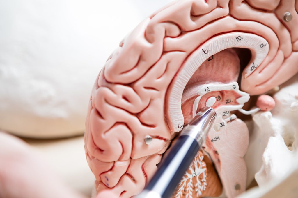
The pineal gland, also called the pinealis, is a small gland in the midbrain area and is located deep inside the skull. It is part of the hormone system of the human body. For example, the pineal gland produces the hormone melatonin, which regulates the day-night rhythm.
Tumors in the area of the pineal gland (= pineal tumor) are almost always benign and range from cysts to meningiomas to pinealocytomas and pinealoblastomas
(= malignant variant of the tumor). In childhood and adolescence, pineal gland cysts are most common.
Tumors of the pineal gland are often characterized by nonspecific symptoms such as headache, nausea, vomiting, double vision and imbalance. Because these cysts are rare, the attending physicians often fail to recognize the connection between the cyst and the symptoms.
A pineal cyst located in the immediate vicinity of the so-called aqueduct (which connects the water chambers of the brain) can cause temporary closure or constriction of the aqueduct. This results in outflow disorders of the liquor (= brain fluid), which can cause the various symptoms.
In addition to a detailed clinical-neurological examination, neuroradiologic imaging by MRI (“tube”) must be performed for the above complaints. Thin-slice MRI images (with a layer thickness of 1 mm or less with contrast media, native and T2 as well as CISS special sequences) are required to visualize the pineal region with different highlighting.

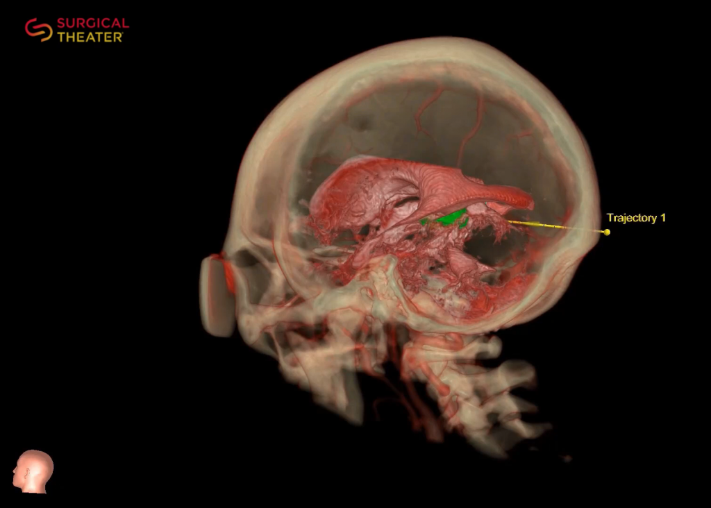
When operating on a pineal cyst or pineal tumor, Prof. Feigl uses minimally invasive endoscopy-supported surgical techniques. In addition to the constant use of intraoperative neuromonitoring (= monitoring of brain and cranial nerve functions), neuronavigation (“GPS for the brain”) is also used. By using special instruments, it is possible to remove the cyst/tumor in this region by means of a mini-craniotomy (= opening of the skull) measuring only about 2 x 2.5 cm.
Understanding Pineal Tumors
Request for Treatment
Request for Treatment

Navigation and Imaging using the Surgical Theater System
The pineal gland, also called the pinealis, is a small gland in the midbrain area and is located deep inside the skull. It is part of the hormone system of the human body. For example, the pineal gland produces the hormone melatonin, which regulates the day-night rhythm.
Tumors in the area of the pineal gland (= pineal tumor) are almost always benign and range from cysts to meningiomas to pinealocytomas and pinealoblastomas (= malignant variant of the tumor). In childhood and adolescence, pineal gland cysts are most common.
Tumors of the pineal gland are often characterized by nonspecific symptoms such as headache, nausea, vomiting, double vision and imbalance. Because these cysts are rare, the attending physicians often fail to recognize the connection between the cyst and the symptoms.
A pineal cyst located in the immediate vicinity of the so-called aqueduct (which connects the water chambers of the brain) can cause temporary closure or constriction of the aqueduct. This results in outflow disorders of the liquor (= brain fluid), which can cause the various symptoms.
In addition to a detailed clinical-neurological examination, neuroradiologic imaging by MRI (“tube”) must be performed for the above complaints. Thin-slice MRI images (with a layer thickness of 1 mm or less with contrast media, native and T2 as well as CISS special sequences) are required to visualize the pineal region with different highlighting.
When operating on a pineal cyst or pineal tumor, Prof. Feigl uses minimally invasive endoscopy-supported surgical techniques. In addition to the constant use of intraoperative neuromonitoring (= monitoring of brain and cranial nerve functions), neuronavigation (“GPS for the brain”) is also used. By using special instruments, it is possible to remove the cyst/tumor in this region by means of a mini-craniotomy (= opening of the skull) measuring only about 2 x 2.5 cm.
Pineal Tumor: briefly explained on YouTube
Request for Treatment
Request for Treatment